Electron density  profiles are easily
obtained by Fourier reconstruction from the measured form factors.
This method by itself can only give relative electron densities because
there is an arbitrary scale factor for each sample.
However, our modeling studies to be described later give
estimates for these scale factors; these are used in the Fourier
electron density profiles in Fig. 6 which are therefore displayed in absolute
units. There are three different samples for which we obtained fourth order
form factors and we have averaged these electron density profiles
using the standard phases
profiles are easily
obtained by Fourier reconstruction from the measured form factors.
This method by itself can only give relative electron densities because
there is an arbitrary scale factor for each sample.
However, our modeling studies to be described later give
estimates for these scale factors; these are used in the Fourier
electron density profiles in Fig. 6 which are therefore displayed in absolute
units. There are three different samples for which we obtained fourth order
form factors and we have averaged these electron density profiles
using the standard phases  . The resulting solid curve in Fig. 6
shows a terminal methyl trough centered at the middle of the bilayer
at z=0 and two headgroup peaks, with head-head separation
. The resulting solid curve in Fig. 6
shows a terminal methyl trough centered at the middle of the bilayer
at z=0 and two headgroup peaks, with head-head separation
 =39.6
=39.6 . For comparison, the
electron density profile obtained from the uncorrected form
factors is shown by a dotted curve in Fig. 6. This latter profile
has (i) wider headgroup peaks and
methyl trough, but (ii) the position of the headgroup peak is
very close to the position of the headgroup peak in the fluctuation
corrected profile shown by the solid curve in Fig. 6. Both
these properties follow from the general theory (Zhang et al., 1994).
These results were also anticipated in earlier studies that postulated
phenomenological Debye-Waller factors (Franks and Lieb, 1979;
Torbet and Wilkins, 1976; Zaccai et al., 1975).
Some details of our derivation and differences with the
preceding ideas are given in Appendix A. The significance
of this result for evaluation of earlier fluctuation uncorrected
analyses of bilayer structure is that estimates of head-head
spacing
. For comparison, the
electron density profile obtained from the uncorrected form
factors is shown by a dotted curve in Fig. 6. This latter profile
has (i) wider headgroup peaks and
methyl trough, but (ii) the position of the headgroup peak is
very close to the position of the headgroup peak in the fluctuation
corrected profile shown by the solid curve in Fig. 6. Both
these properties follow from the general theory (Zhang et al., 1994).
These results were also anticipated in earlier studies that postulated
phenomenological Debye-Waller factors (Franks and Lieb, 1979;
Torbet and Wilkins, 1976; Zaccai et al., 1975).
Some details of our derivation and differences with the
preceding ideas are given in Appendix A. The significance
of this result for evaluation of earlier fluctuation uncorrected
analyses of bilayer structure is that estimates of head-head
spacing  should be reliable, but widths of structural
features such as headgroups will have been overestimated.
Fortunately,
should be reliable, but widths of structural
features such as headgroups will have been overestimated.
Fortunately,  has been the important quantity for
most applications (McIntosh and Simon, 1986a and 1986b).
has been the important quantity for
most applications (McIntosh and Simon, 1986a and 1986b).
Figure 6 also shows the Fourier reconstructions of
the DPPC gel phase electron density profile with 4 orders and
with 10 orders (Torbet and Wilkins, 1976). It is remarkable
that  is essentially the same for both these reconstructions,
especially since it is smaller by 2Å for
is essentially the same for both these reconstructions,
especially since it is smaller by 2Å for  =6 and by 1Å for
 =8 (Wiener et al., 1989). There is also evidence that
this fortuitous accuracy in the h=4 value for
=8 (Wiener et al., 1989). There is also evidence that
this fortuitous accuracy in the h=4 value for  appears to
hold for the fluid phase as well. This evidence comes from
Fourier analyzing the electron density profile obtained from
molecular dynamics simulations (Tu et al., 1995) and reconstructing the
Fouriers for various orders h. The peak position of the
electron density from the simulation is at z=18.3Å and the peak positions
for the Fouriers are 18.9Å (h=2), 19.9Å (h=3), 18.3Å (h=4),
19.2Å (h=5) and 18.6Å (h=6). The difference in
appears to
hold for the fluid phase as well. This evidence comes from
Fourier analyzing the electron density profile obtained from
molecular dynamics simulations (Tu et al., 1995) and reconstructing the
Fouriers for various orders h. The peak position of the
electron density from the simulation is at z=18.3Å and the peak positions
for the Fouriers are 18.9Å (h=2), 19.9Å (h=3), 18.3Å (h=4),
19.2Å (h=5) and 18.6Å (h=6). The difference in  from the
gel phase to the fluid phase will be important in the
next subsection; half this difference
from the
gel phase to the fluid phase will be important in the
next subsection; half this difference  is indicated by the
distance between the vertical dashed lines in Fig. 6.
is indicated by the
distance between the vertical dashed lines in Fig. 6.
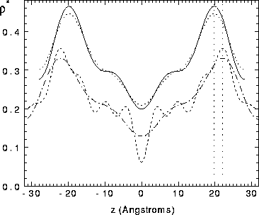
Figure: Electron density profiles  , in
absolute units of electrons/Å
, in
absolute units of electrons/Å , as a function
of z along the bilayer normal with the center of the bilayer at z=0,
obtained by Fourier reconstruction with phases
, as a function
of z along the bilayer normal with the center of the bilayer at z=0,
obtained by Fourier reconstruction with phases  .
Solid line: average from three samples of
.
Solid line: average from three samples of  phase DPPC
using four orders of diffraction.
Dotted line: same average except that
uncorrected form factors were used.
Dash-dot line: gel phase DPPC using four orders, from
Wiener et al. (1989).
Dashed line: gel phase DPPC using ten orders.
To avoid overlapping, the gel phase curves have been displaced downwards by -0.1 electrons/Å
phase DPPC
using four orders of diffraction.
Dotted line: same average except that
uncorrected form factors were used.
Dash-dot line: gel phase DPPC using four orders, from
Wiener et al. (1989).
Dashed line: gel phase DPPC using ten orders.
To avoid overlapping, the gel phase curves have been displaced downwards by -0.1 electrons/Å .
.